Find out more about our range of electron microscopes with specialist detectors operated by specialist trained dedicated staff aimed at a broad range of applications for both routine and urgent analysis reporting.
Focused ion beam field emission scanning electron microscope (FIB-FESEM)

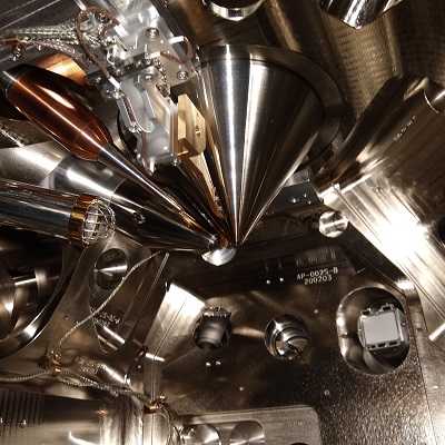
- TESCAN S8215G, focused ion beam field emission scanning electron microscope (FIB-FESEM),
- Ga focused ion beam source,
- Secondary and backscatter electron detectors,
- Oxford Instruments Symmetry EBSD,
- Oxford Instruments Ultim® Max EDS,
- Nanomanipulator for lamella liftouts,
- Pt gas injection system.

Left: gas injection system tip, nanomanipulator tip and Cu grid inside the S8000G sample chamber during TEM lamella production,
Right: FIB milled TEM lamella on a nanomanipulator tip during alignment with the Cu post on TEM grid prior to attachment.
FIB-FESEM with integrated TOF-SIMS

- TESCAN Solaris X, focused ion beam field emission scanning electron microscope (FIB-FESEM) and integrated time-of-flight secondary ion mass spectrometer (TOF-SIMS),
- Field emission electron source and immersion lens,
- Xe plasma source,
- Secondary and backscatter electron detectors,
- Nanomanipulator for lamella liftouts.
Scanning electron microscopy

- TESCAN Vega 3, variable pressure scanning electron microscopes for high resolution imaging (three available),
- Giant and large chambers (GMU and LMU),
- Tungsten filament sources,
- High and low vacuum modes,
- Oxford instruments EDS detectors,
- Secondary and backscatter electron detectors,
- Nordlys Nano EBSD (on GMU).
Hitachi SU5000 field emission scanning electron microscope

- Secondary electron detectors, lower, top and variable pressure,
- 5 segment backscatter detector,
- Conventional and variable pressure modes,
- EDAX TEAM EDS microanalysis system.
Hitachi SU3500 scanning electron microscope

- Tungsten filament,
- Secondary detectors; conventional and variable pressure,
- 5 segment backscatter detector,
- EDAX TEAM EDS microanalysis system,
- Conventional and variable pressure modes.
Additional facilities
- Philips CM20 TEM,
- Bruker D5005 X-ray diffractometers,
- Bruker AFM D3100,
- Bruker DEKTAK,
- Micro Materials NanoTest,
- Spectrometry (Raman, UV-VIS and FTIR),
- Expert sample preparation equipment.
Microscopy services - state-of-the-art facilities
Focused ion beam field emission scanning electron microscope (FIB-FESEM)
- TESCAN S8215G, focused ion beam field emission scanning electron microscope (FIB-FESEM),
- Ga focused ion beam source,
- Secondary and backscatter electron detectors,
- Oxford Instruments Symmetry EBSD,
- Oxford Instruments Ultim® Max EDS,
- Nanomanipulator for lamella liftouts,
- Pt gas injection system.
Left: gas injection system tip, nanomanipulator tip and Cu grid inside the S8000G sample chamber during TEM lamella production,
Right: FIB milled TEM lamella on a nanomanipulator tip during alignment with the Cu post on TEM grid prior to attachment.
FIB-FESEM with integrated TOF-SIMS
- TESCAN Solaris X, focused ion beam field emission scanning electron microscope (FIB-FESEM) and integrated time-of-flight secondary ion mass spectrometer (TOF-SIMS),
- Field emission electron source and immersion lens,
- Xe plasma source,
- Secondary and backscatter electron detectors,
- Nanomanipulator for lamella liftouts.
Scanning electron microscopy
- TESCAN Vega 3, variable pressure scanning electron microscopes for high resolution imaging (three available),
- Giant and large chambers (GMU and LMU),
- Tungsten filament sources,
- High and low vacuum modes,
- Oxford instruments EDS detectors,
- Secondary and backscatter electron detectors,
- Nordlys Nano EBSD (on GMU).
Hitachi SU5000 field emission scanning electron microscope
- Secondary electron detectors, lower, top and variable pressure,
- 5 segment backscatter detector,
- Conventional and variable pressure modes,
- EDAX TEAM EDS microanalysis system.
Hitachi SU3500 scanning electron microscope
- Tungsten filament,
- Secondary detectors; conventional and variable pressure,
- 5 segment backscatter detector,
- EDAX TEAM EDS microanalysis system,
- Conventional and variable pressure modes.
Additional facilities
- Philips CM20 TEM,
- Bruker D5005 X-ray diffractometers,
- Bruker AFM D3100,
- Bruker DEKTAK,
- Micro Materials NanoTest,
- Spectrometry (Raman, UV-VIS and FTIR),
- Expert sample preparation equipment.